
Wisdom Tooth · Impaction Guide
Impacted Wisdom Tooth: Symptoms, Types, Surgery & Treatment in Singapore
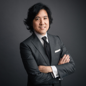 Medically reviewed by Dr Jonathan Liu, BDS (NUS) · SDC Reg. D21395J · 25+ years experience
Last reviewed: June 2026
Medically reviewed by Dr Jonathan Liu, BDS (NUS) · SDC Reg. D21395J · 25+ years experience
Last reviewed: June 2026
The Short Answer
An impacted wisdom tooth is a third molar that cannot fully erupt because the jaw has insufficient space or an adjacent tooth is blocking the path. It may grow sideways, angle forward toward the second molar, or remain completely buried in bone. Not all impacted wisdom teeth require removal — the decision depends on whether the tooth is causing or likely to cause clinical harm, confirmed by OPG X-ray and clinical assessment.
Surgical removal is MediSave-claimable for Singapore Citizens and PRs, with withdrawal limits of S$420–S$2,440 per session depending on case complexity under the MOH Table of Surgical Procedures.
At a Glance
Key Facts About Impacted Wisdom Teeth
Common Age
17–25
When wisdom teeth typically erupt — often without enough space
Prevalence
~9 in 10
Adults have at least one impacted or partially impacted wisdom tooth
Surgery Cost at Dentalis
S$950–S$2,000
Per tooth, before GST · MediSave-claimable
MediSave Limits
S$420–S$2,440
TOSP Table 1B–4B · + S$300 surgical consumables
Recovery
3–7 Days
Initial healing · Swelling peaks Days 2–3 · MC issued same day
First Visit at Dentalis
S$98
Consultation, Examination + OPG · Before GST
Definition
What Is an Impacted Wisdom Tooth?
An impacted wisdom tooth is a third molar that fails to fully erupt into the mouth because of insufficient jaw space or obstruction. It can be partially impacted — breaking partially through the gum — or fully impacted, remaining entirely within the jawbone or covered by gum tissue.
Wisdom teeth are the last permanent teeth to develop, typically emerging between ages 17 and 25. Most adults have four — one at each corner of the dental arches. As human jaws evolved to their current, smaller form, space for third molars reduced. When a tooth cannot move into the correct position, it becomes impacted.
Impacted wisdom teeth are among the most common conditions managed by dentists in Singapore. They are frequently detected on routine OPG panoramic X-rays before any symptoms appear. Some impacted teeth remain stable for years; others progress to infection, decay in the adjacent second molar, or cyst formation — making early assessment important even in the absence of pain.
As noted in guidance from the National Dental Centre Singapore (NDCS): impacted wisdom teeth can cause tooth decay of the wisdom tooth and adjacent second molar, infection and inflammation of overlying gum tissue, and — in more serious cases — cyst formation that damages surrounding jawbone. The absence of pain does not confirm the tooth is safe.
Classification
What Are the Types of Impacted Wisdom Teeth?
Impacted wisdom teeth are classified by two factors: their angle of growth within the jaw, and how deeply they are buried. Both determine symptoms, surgical complexity, imaging requirements, and MediSave table classification.
By angle of impaction
| Type | Position | Clinical Significance | Frequency |
|---|---|---|---|
| Mesioangular | Angled forward toward the second molar | Most common. High risk of food trapping, pericoronitis, and decay on the second molar root surface | Most common |
| Horizontal | Lying completely sideways, parallel to the jawbone | Highest surgical complexity — tooth must be sectioned for removal. Close inferior alveolar nerve proximity common. CBCT often required. | Second most common (lower jaw) |
| Vertical | Upright but unable to fully erupt through gum | May partially emerge. Risk depends on depth of bone coverage over the crown | Common |
| Distoangular | Angled backward toward the rear of the jaw | Less common. Angulation away from normal access can increase surgical difficulty | Least common |
By depth of burial
Soft Tissue Impaction
Tooth has erupted through bone but remains covered by gum tissue only. Gum flap creates pericoronitis risk. Surgical access is simpler than bony impaction.
Partial Bony Impaction
Part of the tooth crown is still embedded in bone. Bone removal is required during surgery. MediSave-claimable. Corresponds to MOH TOSP Table 1B or 2C.
Complete Bony Impaction
Entire tooth remains within the jawbone. May be asymptomatic for years but can develop cysts. Requires the most complex surgical approach. Corresponds to MOH TOSP Table 3A.
Clinical Presentation
What Are the Symptoms of an Impacted Wisdom Tooth?
Many impacted wisdom teeth produce no symptoms at all — and may still be causing damage. The absence of pain does not confirm the tooth is safe. Regular OPG monitoring is the only reliable way to detect silent damage to the adjacent second molar.
When symptoms are present
- Pain or pressure at the back of the mouth, often around the last molar
- Swollen, red, or bleeding gum tissue surrounding the tooth
- Difficulty fully opening the mouth — trismus from jaw muscle inflammation
- Bad taste or persistent bad breath from bacteria under a gum flap
- Jaw swelling or facial tenderness on the affected side
- Headache or earache from referred nerve pressure
- Swollen lymph nodes under the jaw or in the neck
- Pus or discharge near the back gum area
Silent damage — no symptoms
- Decay forming on the root surface of the adjacent second molar — often invisible without X-ray
- Bone loss (periodontal pocket) forming between the wisdom tooth and second molar
- Dentigerous cyst developing around a fully buried tooth crown — may cause no pain until large
- Root resorption of the second molar from sustained pressure
Why this matters: Damage to the second molar from a neglected impacted wisdom tooth can become irreversible. Losing the second molar is a significantly greater clinical problem than removing the original wisdom tooth.
Dentigerous Cyst
A dentigerous cyst forms within the follicular sac surrounding the crown of a fully impacted wisdom tooth. As it enlarges slowly — often over months or years without any symptoms — it resorbs surrounding jawbone and can displace or damage adjacent teeth. On OPG X-ray it appears as a radiolucent shadow around the tooth crown. Left untreated it can grow large enough to weaken the jaw structurally. Early identification through routine imaging allows for straightforward removal alongside the tooth itself, before the cyst reaches a size that requires separate surgical management.
Why It Happens
Why Do Wisdom Teeth Become Impacted?
The primary reason is a structural mismatch between jaw size and the space required for all teeth to erupt. This is not a hygiene or behavioural problem — it is genetic and anatomical.
- Genetic jaw size — smaller modern jaws leave insufficient room for third molars that evolved for larger ancestral jaws
- Late eruption timing — wisdom teeth attempt to emerge after all other permanent teeth are already established and occupying available space
- Abnormal angulation — the tooth develops at an angle that prevents normal upward eruption
- Adjacent tooth obstruction — the second molar blocks the eruption path
- Dense overlying bone — particularly in older patients, where bone is less elastic and eruption against resistance becomes impractical
Impaction cannot be prevented. However, it can be detected early through regular dental check-ups and OPG X-rays — allowing planned management before complications develop. Detection in a patient's early twenties, when root formation is incomplete and bone is more elastic, generally means a simpler surgical procedure than detection at 35 or 40.
The Central Decision
Does an Impacted Wisdom Tooth Always Need to Be Removed?
No. Evidence-based guidance from Cochrane systematic reviews confirms that prophylactic removal of asymptomatic impacted wisdom teeth is not always warranted. A fully impacted, stable tooth that is not causing damage may be monitored rather than removed. The correct decision requires imaging and clinical assessment — not symptoms alone.
| Clinical Finding | Typical Recommendation |
|---|---|
| Fully impacted, symptom-free, stable, not causing damage on imaging | Monitor with periodic OPG — extraction not currently indicated |
| Partially erupted, first episode of pericoronitis, mild | Professional cleaning, antibiotics if indicated, reassess — may monitor or extract |
| Partially erupted, recurrent pericoronitis (2+ episodes) | Extraction recommended — gum flap will not resolve while tooth remains |
| Decay forming on root surface of adjacent second molar | Extraction recommended — second molar at risk of irreversible damage |
| Dentigerous cyst confirmed on imaging | Extraction required — cysts cause progressive bone loss if untreated |
| Mesioangular impaction pressing on second molar root | Extraction recommended — risk of root resorption |
| Horizontal impaction, asymptomatic, no damage | Decision based on age, root development, patient risk profile — discuss at consultation |
At Dentalis, no extraction is recommended without first establishing clear clinical justification visible on imaging. The decision is individual, not algorithmic.
Risks of Delay
What Happens If an Impacted Wisdom Tooth Is Left Untreated?
When a clinically problematic impacted tooth is left without treatment, complications typically worsen over time and become progressively more difficult to manage.
| Complication | What Happens | Reversible? |
|---|---|---|
| Pericoronitis | Recurring bacterial infection of the gum flap around a partially erupted tooth. Each episode may be more severe than the last. Risk of spreading infection to jaw or throat. | Resolves with treatment — recurs while gum flap remains |
| Second molar decay | Food and bacteria accumulate in the contact zone between the impacted tooth and the second molar root. Cavity forms on the second molar without any symptoms. | Cavity treatable if caught early — root damage may be irreversible if late |
| Periodontal bone loss | Chronic bacterial accumulation in the pocket between the two teeth causes progressive bone loss around both the wisdom tooth and the distal root of the second molar. | Bone loss is permanent once established |
| Root resorption of second molar | Sustained pressure from a mesioangular or horizontal impacted tooth causes the roots of the adjacent second molar to dissolve. This is irreversible and may require second molar extraction. | Not reversible — second molar may be lost |
| Dentigerous cyst | A fluid-filled cyst forms around the crown of a fully buried wisdom tooth. Expands silently, eroding surrounding bone. May displace adjacent teeth or damage the inferior alveolar nerve if large. | Bone loss from cyst expansion not fully reversible |
| Higher surgical risk | Root formation completes with age, bone density increases, and surgical access becomes more restricted. A procedure that is straightforward at 22 may be significantly more complex at 40. | Complexity increases with age — early removal is generally simpler |
Imaging & Assessment
How Is an Impacted Wisdom Tooth Diagnosed?
Diagnosis requires clinical examination combined with dental imaging. A clinical examination alone cannot determine impaction angle, depth, root morphology, or proximity to the inferior alveolar nerve — imaging is essential before any treatment decision is made.
OPG X-ray (Orthopantomogram)
A panoramic digital X-ray showing all teeth, bone levels, and surrounding structures in a single image. Standard first-line imaging for assessing impaction type, depth, root number and shape, and the position of the inferior alveolar nerve canal relative to the wisdom tooth roots. At Dentalis, OPG is taken at the first consultation and examination (S$98 before GST) and is reviewed with the patient before any recommendation is made.
CBCT Scan (Cone Beam CT)
Three-dimensional imaging used when the inferior alveolar nerve is in close proximity to the lower wisdom tooth roots on OPG — where a two-dimensional image is insufficient to safely plan the surgical approach. CBCT provides precise spatial data: the exact relationship between the tooth root and the nerve canal in three planes, which determines the surgical technique and whether coronectomy should be considered. At Dentalis, CBCT is available in-clinic (S$180–S$500 before GST) — no external imaging referral required.
The clinical examination assesses gum condition, mouth opening range, lymph node status, jaw movement, and any visible signs of infection, pericoronitis, or adjacent molar decay.
The inferior alveolar nerve runs through the mandible (lower jaw) and provides sensation to the lower lip, chin, gum, and teeth. In cases where an impacted lower wisdom tooth root is close to this nerve, careful pre-surgical imaging — and sometimes coronectomy rather than full extraction — reduces the risk of post-operative nerve paresthesia. CBCT is the appropriate tool when OPG alone does not provide sufficient spatial resolution to make this assessment safely.
Treatment Options
What Treatment Is Available for an Impacted Wisdom Tooth?
| Treatment | Indicated When | What It Involves |
|---|---|---|
| Monitoring | Tooth is fully impacted, symptom-free, and stable on imaging with no damage to adjacent structures. Younger patient with low risk profile. | Periodic OPG X-rays at agreed intervals — typically every 1–2 years — to check for changes in tooth position, bone levels, or cyst development. |
| Surgical removal | Recurrent pericoronitis, decay in second molar, cyst formation, bone loss, or persistent pain. Most partially impacted and many fully impacted teeth. | Gum incision, bone reduction, tooth sectioning where required, extraction, and resorbable sutures. Performed under local anaesthesia with optional IV sedation. |
| Coronectomy | Lower wisdom tooth in very high-risk proximity to the inferior alveolar nerve where full extraction carries unacceptable paresthesia risk. | The crown of the tooth is removed but the roots are left in situ to protect the nerve. Roots may migrate over time and rarely require later removal. |
The majority of clinically indicated impacted wisdom teeth are treated by surgical removal. Coronectomy is reserved for specific high-risk nerve-proximity cases and is determined by CBCT imaging findings, not patient preference.
When Coronectomy Is Considered
Where the roots of a lower wisdom tooth are inseparably close to the inferior alveolar nerve canal, full removal carries a meaningful risk of nerve injury and postoperative paraesthesia. In these cases, Dr Liu assesses whether coronectomy — deliberate removal of the crown only, leaving the roots in situ — is the safer approach.
The retained roots typically become quiescent and do not cause ongoing problems. They may resorb slowly over years, and in a small percentage of cases migrate away from the nerve and require later removal — though this is uncommon.
Coronectomy is not a default option or a way to avoid surgery. It is a clinical decision made only after CBCT imaging confirms the three-dimensional relationship between the root and the nerve canal. If it is the recommended approach, it is discussed fully and quoted separately at consultation.
The Procedure
What Happens During Impacted Wisdom Tooth Surgery?
Surgery is performed under local anaesthesia and typically takes 30–60 minutes per tooth. Most patients leave the clinic within one to two hours of arrival. The procedure is not painful — patients feel pressure and movement, not sharp pain.
-
Assessment and Imaging OPG panoramic X-ray confirms tooth position, root shape, and nerve proximity. Where the inferior alveolar nerve is in close proximity on OPG, CBCT 3D imaging maps the three-dimensional relationship before surgery is planned. Both imaging modalities are available in-clinic at Dentalis — no referral required. The surgical approach is determined before the patient is in the chair.
-
Anaesthesia via Calaject Local anaesthesia is delivered using Calaject — a computer-controlled anaesthetic delivery system that administers solution at a constant, low-pressure flow rate. This significantly reduces the sensation of the injection itself — the moment most patients find most anxiety-inducing — and produces more consistent, reliable anaesthesia throughout the surgical site. IV sedation is available for patients who prefer to be sedated throughout.
-
Surgical Access A small incision is made in the gum tissue overlying the tooth. Overlying bone is carefully reduced using a surgical handpiece to expose the tooth crown. The amount of bone removed is determined by the depth of impaction — this is the primary driver of surgical complexity and MediSave TOSP table classification.
-
Tooth Removal The tooth is elevated from its socket. For complex impactions — horizontal, deep bony, or multi-rooted cases — the tooth is sectioned into two or more pieces to allow safe removal with minimal force on surrounding structures. For nerve-proximity cases, each piece is removed with careful attention to the inferior alveolar nerve position confirmed on pre-operative imaging. Complex nerve-proximity cases at Dentalis are assessed individually and, where indicated, performed by our visiting oral and maxillofacial surgeon — confirmed at consultation.
-
Closure and Discharge The surgical site is irrigated, debrided, and closed with resorbable sutures which dissolve over 7–10 days. A post-operative care plan — including prescribed analgesics, antibiotics where clinically indicated, and explicit aftercare instructions — is provided before discharge. Medical leave is issued on the day. Most patients leave within the hour.
What to Expect
How Long Is Recovery After Impacted Wisdom Tooth Surgery?
Most patients recover within 5–7 days for a single lower surgical removal, with swelling peaking at Days 2–3. A detailed day-by-day recovery guide — covering swelling timeline, dry socket (alveolar osteitis) prevention, what to eat, MC duration, and when to call the clinic — is at our wisdom tooth recovery guide.
Fees at Dentalis
How Much Does Impacted Wisdom Tooth Surgery Cost in Singapore?
| Procedure | Fee (before GST) | Notes |
|---|---|---|
| Consultation and examination | S$98 | Clinical assessment by Dr Jonathan Liu · OPG X-ray included |
| CBCT 3D Scan | S$180–S$500 | For complex nerve-proximity cases · In-clinic at Dentalis |
| Surgical Removal (impacted tooth) | S$950–S$2,000 | MediSave-claimable · Calaject anaesthesia · Sutures included |
| IV Sedation | S$1,200 per hour | Optional · Quoted at consultation |
All prices before 9% GST. An itemised estimate — including MediSave entitlement for qualifying surgical cases — is provided before any procedure is scheduled.
→ Full cost guide — MediSave scenarios, CHAS, public vs private
MediSave Claimable
Can MediSave Be Used for Impacted Wisdom Tooth Surgery?
Yes. MediSave applies to surgical removal of impacted wisdom teeth for Singapore Citizens and PRs. The procedure must be classified under the MOH Table of Surgical Procedures (TOSP). Classification depends on whether bone removal and tooth division are required, and how many teeth are treated in one session. Simple extractions of fully erupted teeth are not MediSave-claimable.
| MOH TOSP Table | Procedure | Limit | + Consumables | Total |
|---|---|---|---|---|
| Table 1B | Bone removal, no tooth division · 1 tooth | S$420 | S$300 | S$720 |
| Table 2C | Bone removal + tooth division · 1 tooth | S$1,120 | S$300 | S$1,420 |
| Table 3A | Completely buried, bone removal + division · 1 tooth | S$1,390 | S$300 | S$1,690 |
| Table 3B–3C | 2–3 teeth | S$1,740–S$1,920 | S$300 | S$2,040–S$2,220 |
| Table 4A–4B | 4 teeth | S$2,380–S$2,440 | S$300 | S$2,680–S$2,740 |
Source: CPF Board MediSave Withdrawal Limits, effective 1 April 2025. TOSP classification confirmed by Dr Liu after clinical assessment and imaging. At Dentalis, MediSave claims are submitted on your behalf.
→ MediSave for wisdom tooth surgery — full guide with out-of-pocket scenarios
"The teeth most worth protecting are the ones next to the problem tooth. An impacted wisdom tooth that is silently pressing against the second molar root can cause irreversible damage before anyone feels a thing. That is why an X-ray matters more than a symptom history — and why I review the imaging carefully before making any recommendation, in either direction."
Dr Jonathan Liu · BDS (NUS) · Principal Dentist, Dentalis · 25+ Years
Common Questions
Frequently Asked Questions — Impacted Wisdom Tooth
A third molar that cannot fully erupt through the gum line because of insufficient jaw space or obstruction. It may grow sideways (horizontal), angle forward (mesioangular), angle backward (distoangular), or remain upright but unable to break through (vertical). It can be partially impacted — partially visible — or fully impacted, remaining entirely within the bone.
No. A fully impacted, stable tooth not causing damage may be monitored. Removal is recommended when there is recurrent infection, decay in the adjacent second molar, cyst formation, confirmed bone loss, or persistent pain affecting daily life. The correct decision requires OPG X-ray and clinical assessment — not symptoms alone.
A problematic impacted wisdom tooth left untreated typically causes progressive complications: recurrent pericoronitis, decay on the adjacent second molar root, periodontal bone loss, root resorption of the second molar, and potentially cyst formation causing jawbone damage. Damage to the second molar can be irreversible — losing it is a significantly greater clinical problem than removing the original wisdom tooth.
Yes. MediSave applies to surgical removal of impacted wisdom teeth for Singapore Citizens and PRs. Withdrawal limits range from S$420 (Table 1B) to S$2,440 (Table 4B) plus up to S$300 for surgical consumables, effective 1 April 2025. The applicable table is confirmed after clinical assessment. At Dentalis, MediSave claims are submitted on your behalf.
Surgical removal costs S$950–S$2,000 per tooth before GST at Dentalis. First-visit consultation, examination with OPG X-ray is S$98 before GST. For MediSave-qualifying cases, a written cost estimate including your MediSave entitlement is provided before any procedure is scheduled. All fees subject to 9% GST.
Most patients recover within 3–7 days for single lower wisdom tooth removal. Swelling peaks at Days 2–3. Return to desk work is typically 3–5 days. Strenuous exercise should be avoided for 5–7 days. Medical leave of 3–5 days is standard. Full bone healing takes 4–6 weeks.
The procedure is not painful. Local anaesthesia eliminates sharp sensation — patients feel pressure and movement, not pain. At Dentalis, Calaject computer-controlled anaesthesia delivery significantly reduces the discomfort of the injection itself. Post-operative soreness, swelling, and jaw stiffness are expected for 2–4 days and managed with prescribed analgesics.
A partially impacted wisdom tooth has broken through the gum but not fully erupted — leaving a gum flap that traps food and bacteria and creates pericoronitis risk. A fully impacted tooth remains entirely within the bone or gum, with no visible crown above the gum line. Fully impacted teeth may be asymptomatic for years but can still develop cysts or cause adjacent tooth damage visible only on X-ray.
Tanjong Pagar · Singapore CBD
Concerned About an Impacted Wisdom Tooth?
An assessment with Dr Jonathan Liu takes 20–30 minutes and includes a clinical examination and OPG X-ray review. You leave with a clear diagnosis, an honest recommendation — and your MediSave entitlement confirmed in writing if removal is indicated.
Consultation, Examination + OPG X-ray S$98 · Before GST · Same-week appointments · MediSave & CHAS accepted
Clinical Sources
References
Clinical data, MediSave limits, and evidence cited in this guide are drawn from the following primary sources.
- Ghaeminia H et al. — Surgical removal versus retention for the management of asymptomatic disease-free impacted wisdom teeth. Cochrane Database of Systematic Reviews, 2020. Evidence basis for the recommendation framework in this guide: monitoring vs extraction for asymptomatic fully impacted wisdom teeth. Source for the finding that prophylactic removal is not always warranted without clinical indication.
- Renton T — Oral Surgery: Part 4. Minimising and Managing Nerve Injuries and Other Complications. British Dental Journal, 2018. Evidence basis for the CBCT imaging protocol and coronectomy decision framework described in the diagnosis and treatment sections of this guide.
- Ministry of Health Singapore — Guide to Dental Treatment Costs (November 2025). TOSP classification criteria and fee benchmarks for impacted wisdom tooth surgery used in the MediSave and cost sections.
- CPF Board — MediSave Withdrawal Limits for Wisdom Tooth Removal (effective 1 April 2025). Source for all TOSP table limits cited in the MediSave section of this guide.
Medical Review & Credentials
BDS (National University of Singapore) · SDC Registration D21395J · 25+ years in general and oral surgery dentistry · Principal Dentist, Dentalis
Reviewed for clinical accuracy against NDCS guidance, MOH Table of Surgical Procedures, CPF Board MediSave withdrawal limits (effective 1 April 2025), and Cochrane systematic review evidence on prophylactic wisdom tooth removal. Last reviewed: June 2026. Next scheduled review: December 2026.
Dentalis is a MediSave-accredited and CHAS-accredited private dental clinic at 52 Craig Rd, Tanjong Pagar, Singapore 089690 — accessible from Maxwell MRT (Thomson-East Coast Line, Exit 3), Tanjong Pagar MRT (East-West Line, EW15), and Outram Park MRT (EW16/NE3/TE17). Recipient of three global awards in 2026: APAC Insider Best Boutique Dental Practice, APAC Insider Patient Care Excellence, and Business Circle Best Boutique Aesthetic & Restorative Dental Practice. Rated 5.0 from 136 Google reviews.
This article is for informational purposes only and does not replace clinical assessment. Individual cases vary — an OPG X-ray and consultation are required for an accurate diagnosis and treatment recommendation.